J Postgrad Med:视网膜小动脉的螺旋形弯曲暗示主动脉缩窄
2018-10-18 MedSci MedSci原创

印度内洛尔Narayana医学院眼科的Shaik A等人近日在J Postgrad Med杂志上发表了一篇文章,他们报告了一例通过视网膜小动脉的螺旋形弯曲进而推测可能是主动脉缩窄的病例。
印度内洛尔Narayana医学院眼科的Shaik A等人近日在J Postgrad Med杂志上发表了一篇文章,他们报告了一例通过视网膜小动脉的螺旋形弯曲进而推测可能是主动脉缩窄的病例。
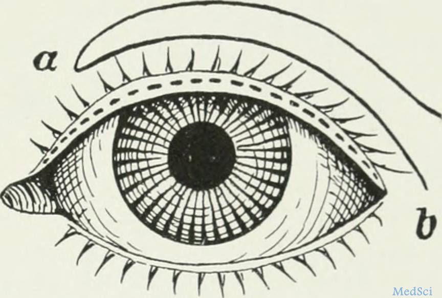
主动脉缩窄(CoA)可以引起阻塞部位上方的血压升高,这种高血压可能被转移并且在视网膜小动脉中有所体现,产生某些高血压性视网膜病变的迹象。进行眼底检查可以有效区分CoA引起的高血压和其他青少年高血压,因为视网膜小动脉的螺旋形仅在CoA中出现,在其他情况下很少出现。
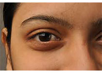
一名16岁高血压男性接受抗高血压治疗,进行常规检查。经检查,双眼的视力均为6/6。双眼的眼底检查显示,在正常的视神经盘中有许多小动脉变窄和加宽的现象。小动脉呈现开瓶器曲折的特征(U形小动脉)。根据眼底检查结果,怀疑可能是CoA并将患者转诊到心脏科进行诊断。在进行超声心动图检查后发现,导管后主动脉缩窄(CoA)。
因此,他们认为,在青少年高血压的病例中,仔细检查眼底可以为全身性疾病诊断提供线索。该病例很好的体现出检眼镜检查在诊断潜在致命全身性疾病中的重要性。
原文出处:
Shaik, A., et al., Corkscrewing of retinal arterioles leading to diagnosis of coarctation of aorta. J Postgrad Med, 2018.
本文系梅斯医学(MedSci)原创编译整理,转载需授权!
本网站所有内容来源注明为“梅斯医学”或“MedSci原创”的文字、图片和音视频资料,版权均属于梅斯医学所有。非经授权,任何媒体、网站或个人不得转载,授权转载时须注明来源为“梅斯医学”。其它来源的文章系转载文章,或“梅斯号”自媒体发布的文章,仅系出于传递更多信息之目的,本站仅负责审核内容合规,其内容不代表本站立场,本站不负责内容的准确性和版权。如果存在侵权、或不希望被转载的媒体或个人可与我们联系,我们将立即进行删除处理。
在此留言


#螺旋形弯曲#
30
#主动脉缩窄#
42
#视网膜#
37
#主动脉#
40
#Med#
0
了解一下,谢谢分享!
76