GUT & Can Res:关新元解析消化道肿瘤研究进展
2012-01-06 MedSci原创 MedSci原创
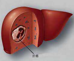
近日, 国际知名杂志GUT和Cancer Research接连刊登了中山大学肿瘤防治中心关新元教授研究团队的最新研究成果,文章中,作者分别对消化道肿瘤中食管癌和肝癌的发病机制进行了探索性研究。 食管癌和肝癌都是严重危害我国人民健康的恶性肿瘤,预后相对差。关新元教授率领研究团队对上述两种肿瘤进行了深入细致的研究工作。分别从肿瘤发生的遗传学基础和肿瘤细胞生长的微环境为立足点对肿瘤的发生机制进行了
近日, 国际知名杂志GUT和Cancer Research接连刊登了中山大学肿瘤防治中心关新元教授研究团队的最新研究成果,文章中,作者分别对消化道肿瘤中食管癌和肝癌的发病机制进行了探索性研究。
食管癌和肝癌都是严重危害我国人民健康的恶性肿瘤,预后相对差。关新元教授率领研究团队对上述两种肿瘤进行了深入细致的研究工作。分别从肿瘤发生的遗传学基础和肿瘤细胞生长的微环境为立足点对肿瘤的发生机制进行了研究。 研究发现,食管鳞状细胞癌的肿瘤相关成纤维细胞能够分泌WNT2促进肿瘤细胞生长,这种效应是通过激活WNT2/beta-catenin通路发挥作用的(GUT 2011, 60(12): 1635-43 )。除环境因素以外,遗传因素在食管癌的发生中也起到重要作用。研究团队发现在食管鳞状细胞癌患者中多数出现抑癌基因RBMS3的表达下调,深入研究发现,该基因可以下调c-Myc,进而影响Rb的磷酸化而发挥抑癌功能,该基因在患者肿瘤组织中的表达情况与患者的预后显著相关,研究结果发表于 Cancer Research 2011, 71(19): 6106-15。
此外,关新元教授研究团队还发现CHD1L与肝癌治疗的化疗耐药有关,并构建了针对CHD1L的腺病毒携带的干扰载体,体内、体外试验结果表明,降低CHD1L表达后能够显著增加肝癌细胞对化疗药物的敏感性,取得很好的疗效(GUT 2011, 60(4): 534-43)。
关新元教授主持的基于实验研究基础之上系列消化道肿瘤研究立足于找到能够对临床的诊断具有指导意义的新的标志物,并为治疗探索新的依据和新的思路、方法。(生物谷Bioon.com)
Wnt2 secreted by tumour fibroblasts promotes tumour progression in oesophageal cancer by activation of the Wnt/β-catenin signalling pathway.
Li Fu1,2, Chunyu Zhang3, Li-Yi Zhang2, Sui-Sui Dong2, Lu-Hui Lu2, Juan Chen2, Yongdong Dai1, Yan Li1, Kar Lok Kong2, Dora L Kwong2, Xin-Yuan Guan1,2
Objectives Interaction between neoplastic and stromal cells plays an important role in tumour progression. It was recently found that WNT2 was frequently overexpressed in fibroblasts isolated from tumour tissue tumour fibroblasts (TF) compared with fibroblasts from non-tumour tissue normal fibroblasts in oesophageal squamous cell carcinoma (OSCC). This study aimed to investigate the effect of TF-secreted Wnt2 in OSCC development via the tumour–stroma interaction. Methods Quantitative PCR, western blotting, immunohistochemistry and immunofluorescence were used to study the expression pattern of Wnt2 and its effect on the Wnt/β-catenin pathway. A Wnt2-secreting system was established in Chinese hamster ovary cells and its conditioned medium was used to study the role of Wnt2 in cell proliferation and invasion. Results Expression of Wnt2 could only be detected in TF but not in OSCC cancer cell lines. In OSCC tissues, Wnt2(+) cells were mainly detected in the boundary between stroma and tumour tissue or scattered within tumour tissue. In this study, Wnt2-positive OSCC was defined when five or more Wnt2(+) cells were observed in 200× microscopy field. Interestingly, Wnt2-positive OSCC (22/51 cases) was significantly associated with lymph node metastases (p=0.001), advanced TNM stage (p=0.001) and disease-specific survival (p<0.0001). Functional study demonstrated that secreted Wnt2 could promote oesophageal cancer cell growth by activating the Wnt/β-catenin signalling pathway and subsequently upregulated cyclin D1 and c-myc expression. Further study found that Wnt2 could enhance cell motility and invasiveness by inducing epithelial–mesenchymal transition. Conclusions TF-secreted Wnt2 acts as a growth and invasion-promoting factor through activating the canonical Wnt/β-catenin signalling pathway in oesophageal cancer cells.
Downregulation of RBMS3 Is Associated with Poor Prognosis in Esophageal Squamous Cell Carcinoma
Yan Li1, Leilei Chen2, Chang-jun Nie1, Ting-ting Zeng1, Haibo Liu1, Xueying Mao1, Yanru Qin3, Ying-Hui Zhu1, Li Fu2, and Xin-Yuan Guan1,2
Deletions on chromosome 3p occur often in many solid tumors, including esophageal squamous cell carcinoma (ESCC), suggesting the existence at this location of one or more tumor suppressor genes (TSG). In this study, we characterized RBMS3 gene encoding an RNA-binding protein as a candidate TSG located at 3p24. Downregulation of RBMS3 mRNA and protein levels was documented in approximately 50% of the primary ESCCs examined. Clinical association studies determined that RBMS3 downregulation was associated with poor clinical outcomes. RBMS3 expression effectively suppressed the tumorigenicity of ESCC cells in vitro and in vivo, including by inhibition of cell growth rate, foci formation, soft agar colony formation, and tumor formation in nude mice. Molecular analyses revealed that RBMS3 downregulated c-Myc and CDK4, leading to subsequent inhibition of Rb phosphorylation. Together, our findings suggest a tumor suppression function for the human RBMS3 gene in ESCC, acting through c-Myc downregulation, with genetic loss of this gene in ESCC contributing to poor outcomes in this deadly disease. 、
Clinical significance of CHD1L in hepatocellular carcinoma and therapeutic potentials of virus-mediated CHD1L depletion
Leilei Chen1,3,4, Yun-Fei Yuan2, Yan Li1, Tim Hon Man Chan3,4, Bo-Jian Zheng5, Jun Huang2, Xin-Yuan Guan1,3,4
Background Hepatocellular carcinoma (HCC) is among the most lethal of human malignancies. It is difficult to detect early, has a high recurrence rate and is refractory to chemotherapies. Amplification of 1q21 is one of the most frequent genetic alterations in HCC. CHD1L is a newly identified oncogene responsible for 1q21 amplification. This study aims to investigate the role of CHD1L in predicting prognosis and chemotherapy response of patients with HCC, its chemoresistant mechanism and whether virus-mediated CHD1L silencing has therapeutic potentials for HCC treatment. Methods The clinical significance of CHD1L in a cohort of 109 HCC cases including 50 cases who received transarterial chemoembolisation treatment was assessed by clinical correlation and Kaplan–Meier analyses. A CHD1L-overexpressing cell model was generated and the mechanism of chemoresistance involving CHD1L was investigated. An adenovirus-mediated silencing method was used to knockdown CHD1L, and its effects on tumorigenicity and chemoresistance were investigated in vivo and in vitro. Results Overexpression of CHD1L was significantly associated with tumour microsatellite formation (p=0.045), advanced tumour stage (p=0.018), overall survival time (p=0.002), overall survival time of patients who received transarterial chemoembolisation treatment (p=0.028) and chemoresistance (p=0.020) in HCC. Interestingly, CHD1L could inhibit apoptosis induced by 5-fluorourail (5-FU) but not doxorubicin. The mechanistic study revealed that the involvement of the Nur77-mediated pathway in chemotherapeutic agent-induced apoptosis can dictate if CHD1L could confer resistance to chemotherapy. Furthermore, an adenoviral vector containing short hairpin RNAs against CHD1L (CHD1L-shRNAs) could suppress cell growth, clonogenicity and chemoresistance to 5-FU. An in vivo study found that CHD1L-shRNAs could inhibit xenograft tumour growth and increase the sensitivity of tumour cells to 5-FU in nude mice. Conclusions This study highlighted for the first time the prognostic value of CHD1L in HCC and the potential application of virus-mediated CHD1L silencing in HCC treatment.
本网站所有内容来源注明为“梅斯医学”或“MedSci原创”的文字、图片和音视频资料,版权均属于梅斯医学所有。非经授权,任何媒体、网站或个人不得转载,授权转载时须注明来源为“梅斯医学”。其它来源的文章系转载文章,或“梅斯号”自媒体发布的文章,仅系出于传递更多信息之目的,本站仅负责审核内容合规,其内容不代表本站立场,本站不负责内容的准确性和版权。如果存在侵权、或不希望被转载的媒体或个人可与我们联系,我们将立即进行删除处理。
在此留言


#解析#
79
#研究进展#
65
#消化道#
67