从视网膜就能看出阿尔兹海默病端倪?
2021-08-31 K.K MedSci原创

眼睛中出现的视网膜斑点与大脑扫描显示的淀粉样蛋白水平较高相关。这一发现表明,非侵入性视网膜成像检测可能有助于检测早期阿尔兹海默的生物标志物。
淀粉样斑块是由β淀粉样蛋白异常沉积于脑神经元外构成的斑块结构,它阻碍一些功能并最终导致神经元死亡,这也被认为是阿尔茨海默病(AD)的一个标志。但是淀粉样蛋白沉积也可能发生在眼睛的视网膜上,且经常发生在临床诊断为患有阿尔兹海默的病人身上。
在一项小型的横断面研究中,由加州大学圣地亚哥分校医学院的科学家领导的研究小组比较了两项研究中的患者视网膜和大脑淀粉样蛋白的测试,评估淀粉样蛋白水平低的人神经变性风险有多大。研究人员观察到,眼睛中出现的视网膜斑点与大脑扫描显示的淀粉样蛋白水平较高相关。这一发现表明,非侵入性视网膜成像检测可能有助于检测早期阿尔兹海默的生物标志物。该研究结果发表在2021年8月17日的《阿尔茨海默病与痴呆症》杂志上。
Jennifer Ngolab et al, Feasibility study for detection of retinal amyloid in clinical trials: The Anti‐Amyloid Treatment in Asymptomatic Alzheimer's Disease (A4) trial, Alzheimer's & Dementia: Diagnosis, Assessment & Disease Monitoring (2021). DOI: 10.1002/dad2.12199
与淀粉样蛋白风险和神经退行性的纵向评估试验的个体相比,这项试验的四个个体在视网膜上表现出更多的姜黄素阳性点,两组之间存在明显的差异,以前对视网膜中姜黄素标记的淀粉样蛋白的研究已经发现了临床上的患者在早期就表现出认知障碍,临床前队列的视网膜研究则观察到了视网膜包涵体。研究人员证明了在淀粉样蛋白正电子发射断层成像中阳性的无症状个体中可以检测到视网膜姜黄素阳性点升高。
姜黄素在体内与淀粉样蛋白结合,并被用作造影剂来识别早期认知障碍(MCI)以及轻度至中度阿尔兹海默的个体视网膜中的淀粉样蛋白沉积。最近一项使用视网膜眼底成像系统的研究报告称,在MCI和AD的个体中,视网膜斑点的区域性增加,表明区域性淀粉样蛋白沉积可能代表着疾病的发展。此外,视网膜成像系统的成本可能相对较小。
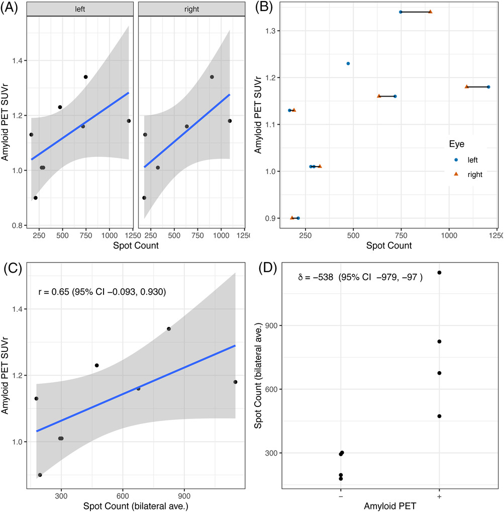
视网膜淀粉样蛋白与脑淀粉样蛋白的比较
加州大学圣地亚哥分校医学院神经科学教授Robert Rissman博士说:"但这些发现是令人鼓舞的,因为它们表明有可能利用视网膜成像检测来确定阿尔茨海默病的发病、扩散和形态,而不是通过昂贵的大脑扫描。我们期待看到不通时间点视网膜扫描的结果,以及Solanezumab单克隆抗体对视网膜成像的影响。不幸的是,我们将需要等待这项试验完成后看到并分析这些数据"。Solanezumab通过结合血液和大脑中的可溶性Aβ蛋白,促使Aβ蛋白在大脑中生成不可溶性的寡聚体和淀粉样斑之前被清除。Solanezumab被用于轻度阿兹海默症和有前驱症状的阿兹海默症的潜在治疗。
Rissman说,下一步将进行更大规模的研究,以更全面地记录和确定视网膜淀粉样蛋白和大脑淀粉样蛋白之间的关系,包括横断面研究。
参考资料:
本网站所有内容来源注明为“梅斯医学”或“MedSci原创”的文字、图片和音视频资料,版权均属于梅斯医学所有。非经授权,任何媒体、网站或个人不得转载,授权转载时须注明来源为“梅斯医学”。其它来源的文章系转载文章,或“梅斯号”自媒体发布的文章,仅系出于传递更多信息之目的,本站仅负责审核内容合规,其内容不代表本站立场,本站不负责内容的准确性和版权。如果存在侵权、或不希望被转载的媒体或个人可与我们联系,我们将立即进行删除处理。
在此留言


#视网膜#
85
#阿尔兹海默#
95
👍
128