彩色多普勒超声诊断右冠状动脉右房瘘合并右房内冠状动脉瘤1例
2019-12-13 王南 任卫东 临床超声医学杂志

患者男,28岁,因心悸于外院超声检查发现右房基底部于上腔静脉入口处见44mm×41mm囊性无回声区。经我院心脏超声心动图检查:全心大,右冠状动脉扩张(图1),根部宽约20mm,自前向后上走行至右房顶部,进入右房内形成局部较大瘤样回声,大小约43mm×41mm;CDFI示主动脉内血流自主动脉右窦经右冠状动脉瘘入右房,在右房内形成瘤样扩张,其内形成环状血流并在其上方可见一破口,约7mm(图2);脉冲多
患者男,28岁,因心悸于外院超声检查发现右房基底部于上腔静脉入口处见44mm×41mm囊性无回声区。经我院心脏超声心动图检查:全心大,右冠状动脉扩张(图1),根部宽约20mm,自前向后上走行至右房顶部,进入右房内形成局部较大瘤样回声,大小约43mm×41mm;CDFI示主动脉内血流自主动脉右窦经右冠状动脉瘘入右房,在右房内形成瘤样扩张,其内形成环状血流并在其上方可见一破口,约7mm(图2);脉冲多普勒于冠状动脉内探及全心动周期高速右冠状动脉至右房分流信号,分流峰速约4.2m/s。超声诊断:右冠状动脉-右房瘘;右房内冠状动脉呈瘤样扩张。
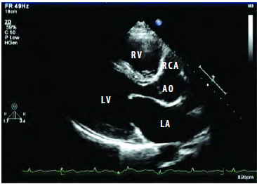
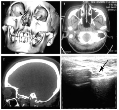
图1 左室长轴切面示右冠状动脉起始部扩张(RV:右室;RCA:右冠状动脉;AO:主动脉;LV:左室;LA:左房)

图2CDFI示右冠状动脉远端于右房内形成瘤样扩张,其上可见破口(RV:右室;RA:右房;CAA:冠状动脉瘤;LA:左房)
CTA检查:右冠状动脉瘤样扩张,最大内径47mm×46mm,瘘口最大横径约10mm(图3)。CTA诊断:右优势型冠状动脉;右冠状动脉扩张,与右房相连。
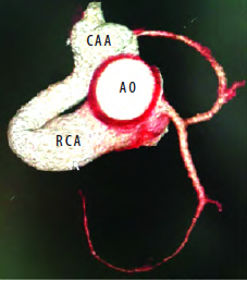
图3 CTA示扩张的右冠状动脉与右房相连,并形成瘤样扩张(AO:主动脉;CAA:冠状动脉瘤;RCA:右冠状动脉)
讨论:
冠状动脉瘘是罕见的先天性心脏病,为冠状动脉的主干或其分支与某心腔或血管的异常通道,病因不明,大多认为在心脏原始发育的过程中,心肌窦状间隙逐渐退化变细形成Thebesion静脉,当某种原因导致心肌间小梁部窦状间隙不退化而持续存在时,即可形成冠状动脉瘘。
本例患者起初因心悸于外院误诊为心脏占位,原因可能是未找到右房内异常血流的起源,只有追踪到异常血流的起源和走行才能做出准确诊断。冠状动脉瘘合并冠状动脉瘤极为罕见,临床表现除出现劳力性呼吸困难和乏力,还可出现冠状动脉痉挛、冠状动脉瘤破裂、冠状动脉瘤血栓及栓塞。冠状动脉瘤无论是单纯性还是继发于冠状动脉瘘,一经确诊均需手术治疗。
超声可以探测到扩张的冠状动脉和瘘入部位的环形无回声区;CDFI可以探测异常血流的起源、走行及瘘入部位,同时可以观察冠状动脉内径的变化及有无血栓生成及狭窄,为后续治疗提供可靠依据。
原始出处:
王南,任卫东.彩色多普勒超声诊断右冠状动脉右房瘘合并右房内冠状动脉瘤1例[J].临床超声医学杂志,2018,20(02):81.
版权声明:
本网站所有内容来源注明为“梅斯医学”或“MedSci原创”的文字、图片和音视频资料,版权均属于梅斯医学所有。非经授权,任何媒体、网站或个人不得转载,授权转载时须注明来源为“梅斯医学”。其它来源的文章系转载文章,或“梅斯号”自媒体发布的文章,仅系出于传递更多信息之目的,本站仅负责审核内容合规,其内容不代表本站立场,本站不负责内容的准确性和版权。如果存在侵权、或不希望被转载的媒体或个人可与我们联系,我们将立即进行删除处理。
在此留言
本网站所有内容来源注明为“梅斯医学”或“MedSci原创”的文字、图片和音视频资料,版权均属于梅斯医学所有。非经授权,任何媒体、网站或个人不得转载,授权转载时须注明来源为“梅斯医学”。其它来源的文章系转载文章,或“梅斯号”自媒体发布的文章,仅系出于传递更多信息之目的,本站仅负责审核内容合规,其内容不代表本站立场,本站不负责内容的准确性和版权。如果存在侵权、或不希望被转载的媒体或个人可与我们联系,我们将立即进行删除处理。
在此留言


#彩色多普勒超声#
66
#多普勒超声#
57
#多普勒#
61
#冠状动脉瘤#
69
#超声诊断#
75
#超声诊断#
53
学习
121