PLoS ONE:肿瘤坏死因子调控褪黑素合成分泌
2012-07-10 Beyond 生物谷
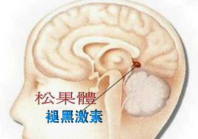
松果体属于室周器官,对机体的防御反应起着关键作用,松果体损伤会引起褪黑激素被抑制。褪黑激素(Melatonin)主要是由哺乳动物和人类的松果体产生的一种胺类激素。近年来,国内外对褪黑激素的生物学功能,尤其是作为膳食补充剂的保健功能进行了广泛的研究,表明其具有促进睡眠、调节时差、抗衰老、调节免疫、抗肿瘤等多项生理功能。 先前已有研究表明体外培养的松果体表达Toll样受体4(TLR4)和肿瘤坏死
松果体属于室周器官,对机体的防御反应起着关键作用,松果体损伤会引起褪黑激素被抑制。褪黑激素(Melatonin)主要是由哺乳动物和人类的松果体产生的一种胺类激素。近年来,国内外对褪黑激素的生物学功能,尤其是作为膳食补充剂的保健功能进行了广泛的研究,表明其具有促进睡眠、调节时差、抗衰老、调节免疫、抗肿瘤等多项生理功能。

先前已有研究表明体外培养的松果体表达Toll样受体4(TLR4)和肿瘤坏死因子受体1(TNFR1),当接受脂多糖(LPS)的刺激时能产生肿瘤坏死因子。近日PLoS ONE杂志上的一项研究目评估了是否是存在于松果体中的星形胶质细胞、小胶质细胞或松果体细胞产生肿瘤坏死因子,以了解松果体活动、褪黑激素的生产与免疫功能之间的相互作用。
实验中用LPS刺激体外培养的松果腺或松果体,肿瘤坏死因子含量的测定采用酶联免疫吸附试验,TLR4和TNFR1的表达用共聚焦显微镜进行分析,免疫组化分析小胶质细胞形态。
在本研究中,数据表明尽管松果腺的主要细胞类型(松果体细胞、星形胶质细胞和小胶质细胞)表达TLR4,但LPS诱导肿瘤坏死因子的生产由小胶质细胞介导。这种效果是由于核因子kappa B(NF-KB)的活化带来的。
此外,研究人员还观察到脂多糖激活小胶质细胞和调节松果体TNFR1的表达。由于肿瘤坏死因子已经证明与炎症反应的放大和延长有关,松果体小胶质细胞产生肿瘤坏死因子表明胶质松果腺细胞网络能调节褪黑激素的释放。这项研究有助从分子与细胞水平上了解在先天免疫反应中褪黑激素是如何调节合成的,研究结果再次证实松果体在免疫反应中充当传感器的作用。

doi:10.1371/journal.pone.0040142
PMC:
PMID:
Glia-Pinealocyte Network: The Paracrine Modulation of Melatonin Synthesis by Tumor Necrosis Factor (TNF)
Sanseray da Silveira Cruz-Machado, Luciana Pinato, Eduardo Koji Tamura, Cláudia Emanuele Carvalho-Sousa,
The pineal gland, a circumventricular organ, plays an integrative role in defense responses. The injury-induced suppression of the pineal gland hormone, melatonin, which is triggered by darkness, allows the mounting of innate immune responses. We have previously shown that cultured pineal glands, which express toll-like receptor 4 (TLR4) and tumor necrosis factor receptor 1 (TNFR1), produce TNF when challenged with lipopolysaccharide (LPS). Here our aim was to evaluate which cells present in the pineal gland, astrocytes, microglia or pinealocytes produced TNF, in order to understand the interaction between pineal activity, melatonin production and immune function. Cultured pineal glands or pinealocytes were stimulated with LPS. TNF content was measured using an enzyme-linked immunosorbent assay. TLR4 and TNFR1 expression were analyzed by confocal microscopy. Microglial morphology was analyzed by immunohistochemistry. In the present study, we show that although the main cell types of the pineal gland (pinealocytes, astrocytes and microglia) express TLR4, the production of TNF induced by LPS is mediated by microglia. This effect is due to activation of the nuclear factor kappa B (NF-kB) pathway. In addition, we observed that LPS activates microglia and modulates the expression of TNFR1 in pinealocytes. As TNF has been shown to amplify and prolong inflammatory responses, its production by pineal microglia suggests a glia-pinealocyte network that regulates melatonin output. The current study demonstrates the molecular and cellular basis for understanding how melatonin synthesis is regulated during an innate immune response, thus our results reinforce the role of the pineal gland as sensor of immune status.
本网站所有内容来源注明为“梅斯医学”或“MedSci原创”的文字、图片和音视频资料,版权均属于梅斯医学所有。非经授权,任何媒体、网站或个人不得转载,授权转载时须注明来源为“梅斯医学”。其它来源的文章系转载文章,或“梅斯号”自媒体发布的文章,仅系出于传递更多信息之目的,本站仅负责审核内容合规,其内容不代表本站立场,本站不负责内容的准确性和版权。如果存在侵权、或不希望被转载的媒体或个人可与我们联系,我们将立即进行删除处理。
在此留言


#分泌#
71
#Plos one#
91
#坏死#
79
#肿瘤坏死因子#
83