PLoS ONE:血管紧张素II(AngII)促进乳腺癌细胞转移
2012-04-25 Deepblue 生物谷
乳腺癌是女性排名第一的常见恶性肿瘤,其癌细胞转移是引起患者死亡的主要原因。目前的研究正在进一步阐明癌症转移的限速步骤,比如循环肿瘤细胞的外渗及侵袭。 血管紧张素II是一种主要的血管活性肽,它局部产生并释放到血液。近日,法国巴黎科尚研究所的研究人员着手完成了一项有意义的研究,他们发现血管紧张素II能够促进癌细胞迁移。 在该研究中,他们使用了一个癌症转移的体内模型。为了研究癌细

乳腺癌是女性排名第一的常见恶性肿瘤,其癌细胞转移是引起患者死亡的主要原因。目前的研究正在进一步阐明癌症转移的限速步骤,比如循环肿瘤细胞的外渗及侵袭。
血管紧张素II是一种主要的血管活性肽,它局部产生并释放到血液。近日,法国巴黎科尚研究所的研究人员着手完成了一项有意义的研究,他们发现血管紧张素II能够促进癌细胞迁移。
在该研究中,他们使用了一个癌症转移的体内模型。为了研究癌细胞转移的重要步骤,他们将能够稳定产生荧光的乳腺癌细胞系(D3H2LN)注入到裸鼠。通过实时活体影像学研究,他们发现血管紧张素II促进了转移灶的形成。
实验发现,用这种多肽预处理后,发生癌细胞转移的老鼠数量增加,并且每个老鼠体内的转移瘤的数量及大小也相应的增加和增大。在体外,血管紧张素II通过促进癌细胞黏附于内皮细胞、跨内皮迁移并通过细胞外基质,促进了癌症转移。
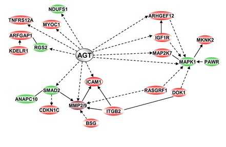
在分子水平,由DNA微阵列分析表明,血管紧张素II预处理后共有102个基因差异性表达。血管紧张素II调控了两类与其前体血管紧张素原紧密相关的基因。在这些基因中,MMP2/MMP9和ICAM1M的上调表达跟由参与细胞黏附、迁移及侵袭的基因组成的网络相关联。
这些结果表明,靶向血管紧张素II可能会成为抑制浸润性乳腺癌转移的有效策略。相关论文发表在4月20日的PLoS ONE。

doi: 10.1371/journal.pone.0035667
PMC:
PMID:
Angiotensin II Facilitates Breast Cancer Cell Migration and Metastasis
Sylvie Rodrigues-Ferreira, Mohamed Abdelkarim, Patricia Dillenburg-Pilla, Anny-Claude Luissint, Anne di-Tommaso, Frédérique Deshayes, Carmen Lucia S. Pontes, Angie Molina, Nicolas Cagnard, Franck Letourneur, Marina Morel, Rosana I. Reis, Dulce E. Casarini, Benoit Terris, Pierre-Olivier Couraud, Claudio M. Costa-Neto, Mélanie Di Benedetto, Clara Nahmias.
Breast cancer metastasis is a leading cause of death by malignancy in women worldwide. Efforts are being made to further characterize the rate-limiting steps of cancer metastasis, i.e. extravasation of circulating tumor cells and colonization of secondary organs.In this study, we investigated whether angiotensin II, a major vasoactive peptide both produced locally and released in the bloodstream, may trigger activating signals that contribute to cancer cell extravasation and metastasis.We used an experimental in vivo model of cancer metastasis in which bioluminescent breast tumor cells (D3H2LN) were injected intra-cardiacally into nude mice in order to recapitulate the late and essential steps of metastatic dissemination. Real-time intravital imaging studies revealed that angiotensin II accelerates the formation of metastatic foci at secondary sites.Pre-treatment of cancer cells with the peptide increases the number of mice with metastases, as well as the number and size of metastases per mouse. In vitro, angiotensin II contributes to each sequential step of cancer metastasis by promoting cancer cell adhesion to endothelial cells, trans-endothelial migration and tumor cell migration across extracellular matrix.At the molecular level, a total of 102 genes differentially expressed following angiotensin II pre-treatment were identified by comparative DNA microarray. Angiotensin II regulates two groups of connected genes related to its precursor angiotensinogen.
本网站所有内容来源注明为“梅斯医学”或“MedSci原创”的文字、图片和音视频资料,版权均属于梅斯医学所有。非经授权,任何媒体、网站或个人不得转载,授权转载时须注明来源为“梅斯医学”。其它来源的文章系转载文章,或“梅斯号”自媒体发布的文章,仅系出于传递更多信息之目的,本站仅负责审核内容合规,其内容不代表本站立场,本站不负责内容的准确性和版权。如果存在侵权、或不希望被转载的媒体或个人可与我们联系,我们将立即进行删除处理。
在此留言


#Plos one#
76
#癌细胞#
69
#细胞转移#
105
#血管紧张素II#
88
#血管紧张素#
94
#癌细胞转移#
78