
J Periodontal Res:L-PRF和A-PRF对牙周成纤维细胞体外伤口愈合的影响
2020-04-30 网络 网络

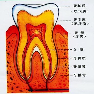
L-PRF和A-PRF+是用于牙周再生手术的自体材料,均来源于患者的血液,但特点不同,对牙周组织成纤维细胞的影响存在争议。
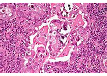
L-PRF和A-PRF+是用于牙周再生手术的自体材料,均来源于患者的血液,但有不同的特点,对牙周组织成纤维细胞的影响存在争议。本研究旨在测定白细胞-富血小板纤维蛋白(L-PRF)和改良型富血小板纤维蛋白(A-PRF+)在体外诱导牙周成纤维细胞增殖和迁移的能力是否存在差异。
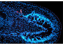
从8名患者中制备L-PRF和A-PRF+膜,在3 mL培养基中培养2天。从7个供体中提取牙龈成纤维细胞(G-F)和牙周膜成纤维细胞(PDL-F)原代细胞。这些细胞在伤口愈合实验板中预培养1天,细胞浓度为3.3×10⁵细胞/mL,留下500±50μm的间隙。将每70ul的细胞悬浮液放入相应的孔中。将预培养的L-PRF和A-PRF+上清液加入其中,并将成纤维细胞再培养24 h,单独培养基(NEG)和成纤维细胞生长因子II(FGF)作为对照。随后,用活细胞成像技术检测24小时内,在37℃、5%CO2条件下细胞的迁移情况。检测细胞增殖和细胞活力。
实验结果:与FGF和NEG相比,在L-PRF和A-PRF+诱导下,细胞增殖速度更快。与对照组相比,A-PRF +和L-PRF诱导的人工伤口愈合速度明显快于对照组。在初始阶段,两种PRF组均诱导了较快的细胞迁移(P < .01),但在停止阶段,A-PRF+组比L-PRF组诱导的迁移率更高(P < .01)。
因此,L-PRF和A-PRF+对牙周成纤维细胞的迁移和增殖均有促进作用,并且,A-PRF+对人工伤口愈合的维持作用比L-PRF久。
原文出处:
Luciano Pitzurra, Effects of L‐PRF and A‐PRF+ on periodontal fibroblasts in in vitro wound healing experiments, journal of periodontal research, 2020, doi.org/10.1111/jre.12714
本网站所有内容来源注明为“梅斯医学”或“MedSci原创”的文字、图片和音视频资料,版权均属于梅斯医学所有。非经授权,任何媒体、网站或个人不得转载,授权转载时须注明来源为“梅斯医学”。其它来源的文章系转载文章,或“梅斯号”自媒体发布的文章,仅系出于传递更多信息之目的,本站仅负责审核内容合规,其内容不代表本站立场,本站不负责内容的准确性和版权。如果存在侵权、或不希望被转载的媒体或个人可与我们联系,我们将立即进行删除处理。
在此留言


#PE#
51
#L-PRF#
63
#成纤维细胞#
72
#伤口愈合#
64
#牙周#
49
#纤维细胞#
48
#PRF#
44